Cytokine Signaling and Allergy-Induced Mental Fatigue
Immune molecules from allergies cross into the brain and directly impair focus and memory.
The cotton-headed, slow-thinking feeling that shows up during allergy season follows a specific, traceable biological process. It follows a specific chain of immune events that ends in the brain, with effects on memory, focus, and processing speed that can be traced back to a molecule-by-molecule cause. Most people file that fog under stress or bad sleep, mostly because the connection between a stuffy nose and a slow brain seems like a stretch. But what if the fog is not a side effect of allergies at all, and instead a direct, mechanical output of the same immune response that causes the sneezing?
Allergy sufferers tend to notice the parts of an allergic reaction that are easiest to see: sneezing, itchy eyes, a congested nose. Fewer people connect that same reaction to slowed thinking or a kind of mental heaviness that sets in during peak pollen weeks. Yet seasonal allergy sufferers report measurable drops in work performance and mental clarity during those weeks, and the fog tends to lift once the season passes, which is itself a clue that something biological, not psychological, is driving it. Indoor allergens like dust mites, mold, and pet dander complicate the picture further, because they operate year-round. Someone exposed to those triggers constantly never gets a clear "before" period to compare against, so the fog just feels like a permanent personality trait rather than a symptom.
That misattribution carries a real cost. When brain fog gets treated as a stress problem or a sleep problem, the treatment aims at the wrong system entirely, and the underlying immune trigger keeps firing. Understanding the actual mechanism, starting with what happens in the seconds after allergen exposure, makes it possible to treat the fog as what it is: a neurological consequence with an identifiable starting point.
IgE Activation and the Cascade That Reaches the Brain
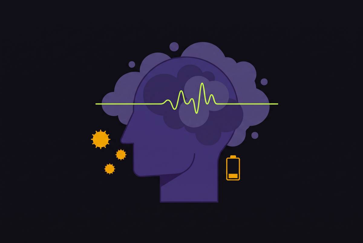
The chain starts in the immune system, not the brain, and it starts fast. When the immune system encounters a recognized allergen, whether that's ragweed pollen or a protein shed from pet dander, IgE antibodies already bound to mast cells trigger a process called degranulation. Degranulation is the rapid release of histamine and other pre-made inflammatory mediators, followed a bit later by the manufacturing and release of inflammatory cytokines built fresh for the occasion.
Key molecules released at this stage include interleukin-6 (IL-6) and tumor necrosis factor-alpha (TNF-α). Both appear in research on sickness behavior (the fatigue and mental dullness that come with a cold or flu), in major depression, and in sleep deprivation. It means the same molecules produced by a ragweed reaction are chemically identical to the ones associated with depressive symptoms and the fog of a sleepless night, a detail that matters because it ties allergic inflammation directly to depressive biochemistry. Elevated IL-6, TNF-alpha, and C-reactive protein appear in both allergic conditions and major depressive disorder, a biochemical overlap.
The IgE-mast cell reaction is the trigger for everything that follows. Cytokine migration into the brain, activation of the brain's immune cells, and disruption of neurotransmitters all follow downstream of that one initiating event. None of it starts in the head. It starts in tissue, with an antibody doing its job a little too aggressively, and from there the question becomes how molecules built for a local, peripheral response end up affecting an organ that is supposed to be walled off from the rest of the body.
Cytokines, the Blood-Brain Barrier, and the Brain's Immune Cells
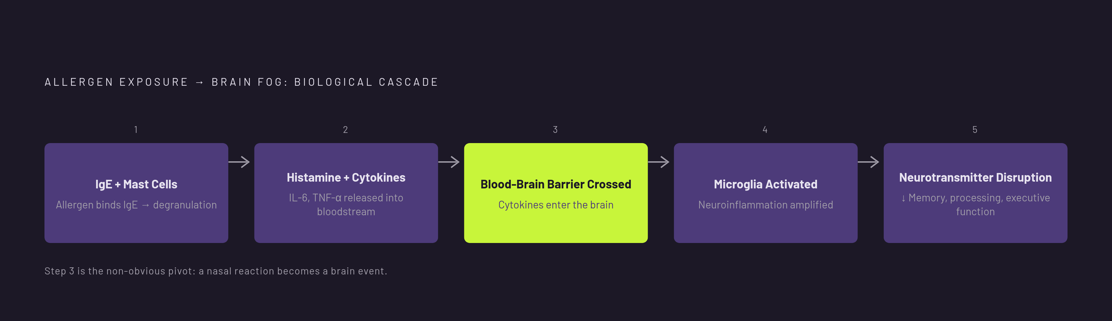
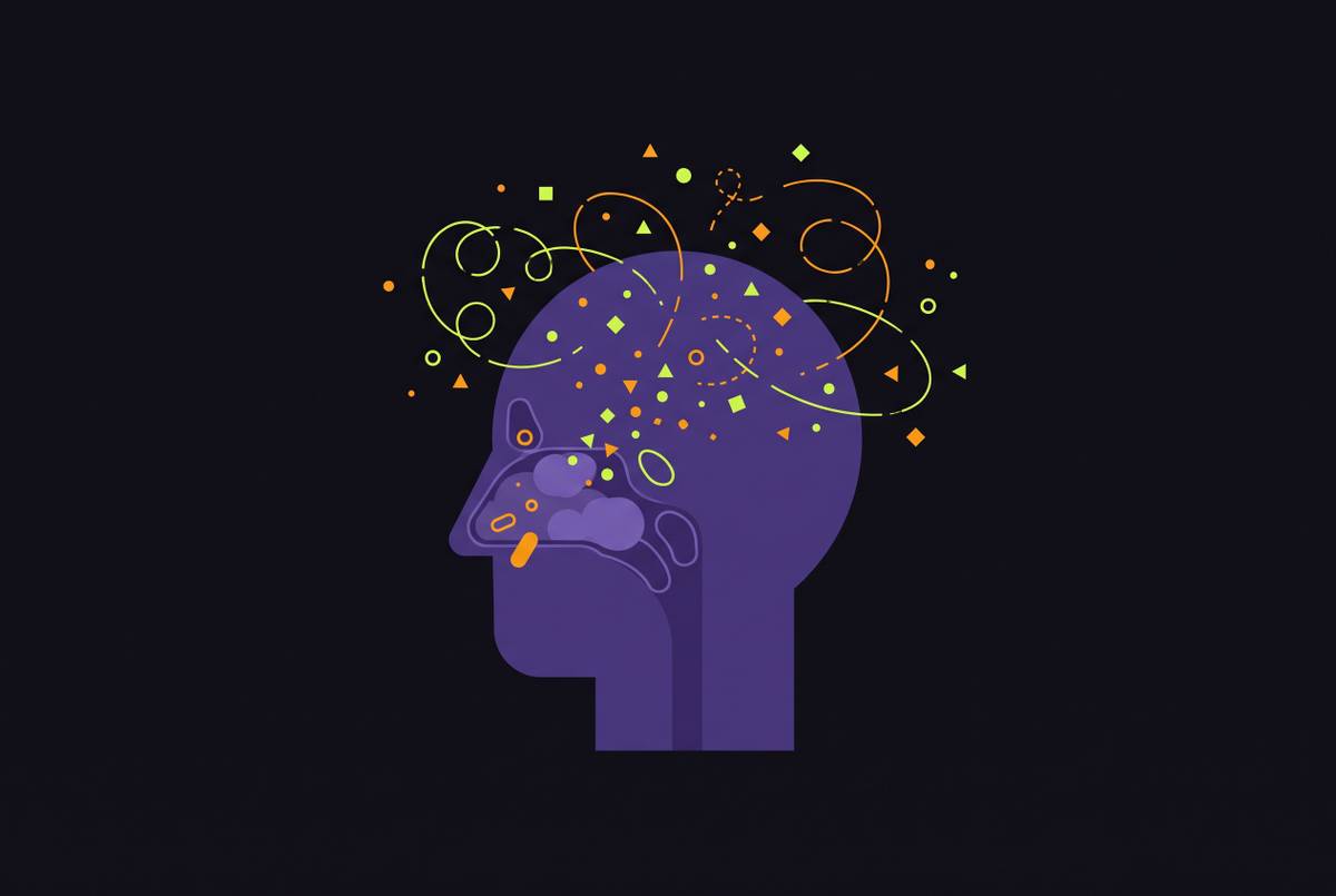
The blood-brain barrier gets described, often, as an impenetrable wall. That description overstates the case. It behaves more like a filter with selective openings, and some of the molecules produced during an allergic reaction happen to fit through.
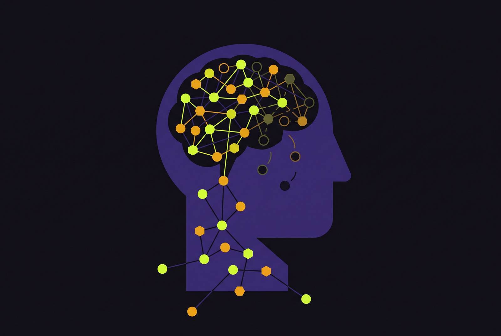
Proinflammatory cytokines, including IL-1β and TNF-α, cross the blood-brain barrier and interfere with the brain's normal communication pathways, reaching neural tissue directly rather than staying confined to the blood. Once those cytokines are inside, they activate microglia, the brain's resident immune cells. Microglia respond to that activation the way immune cells anywhere in the body respond to a threat: by releasing their own inflammatory signals, which amplifies the original peripheral response rather than containing it.
This is the step that answers the question a lot of allergy sufferers ask without realizing it has an answer: why does a nasal reaction make the brain feel slow? Because the same cytokines causing the congestion are, at the same time, gaining direct access to brain tissue and triggering a parallel immune reaction inside it. Neuroimaging work has found decreased activity in prefrontal cortex regions responsible for executive function during active allergic responses, which places a specific, visible signature on what would otherwise be an invisible process. The fog is a change in brain activity, not merely a feeling people are imagining. It is a change in brain activity that can be seen on a scan.
What neuroinflammation does to neurotransmitter balance and cognitive performance
Once cytokines are active in brain tissue, they interfere with the neurotransmitter systems responsible for attention, memory, mood, and processing speed, and the deficits patients describe line up closely with which systems get hit.
Histamine plays a double role here, split between two distinct receptor effects. H1 receptors in the brain mainly drive drowsiness and reduced alertness. H3 receptors regulate the release of dopamine, norepinephrine, and acetylcholine, the neurotransmitters most responsible for focus, memory, and mental clarity. So the same histamine flooding the sinuses during a reaction is also acting directly on receptors that govern how sharp or foggy someone feels.
Proinflammatory cytokines add another layer by disrupting neurotransmitter pathways tied to mood and motivation. IL-6 and TNF-alpha, the same cytokines elevated in major depressive disorder, create a brain chemistry environment that raises the odds of depressive symptoms, independent of how bothered someone feels by their runny nose or watery eyes. A person could rate their physical allergy symptoms as mild, independent of how bothered they feel by their runny nose or watery eyes, and still be dealing with a biochemical environment that pushes toward low mood and mental fatigue.
Chronic allergic stress adds a slower-acting third pathway through cortisol. Ongoing immune activation triggers the HPA axis, raising cortisol, and sustained cortisol elevation impairs the hippocampus's ability to consolidate and retrieve memories. That pathway runs alongside the direct cytokine effects rather than replacing them, so someone dealing with chronic allergies is stacking at least three separate mechanisms working against clear thinking at once.
Put together, the resulting cognitive profile has a specific shape. It appears as reduced processing speed, weaker working memory, and trouble planning or making decisions. Each of those maps to a named mechanism above: processing speed to the direct cytokine interference with brain communication, working memory to cortisol's effect on the hippocampus, and executive function to the same prefrontal disruption visible on imaging.
How disrupted sleep amplifies the neurological damage already underway
Allergic inflammation does not stop once it has disrupted neurotransmitters. It also degrades the sleep the brain depends on to clear out inflammatory waste and consolidate memory, and that creates a second pathway of damage running alongside the first.
Histamine released during an allergic reaction swells the tissue lining the sinuses and nasal passages, which restricts airflow and pushes people toward mouth breathing at night. Mouth breathing fragments sleep architecture and cuts down on time spent in deep, restorative sleep. Coughing and sneezing add a second source of nighttime interruption on top of that.
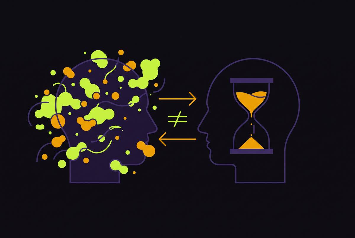
Poor sleep independently raises proinflammatory cytokine levels, which is what turns this into a loop rather than a one-time hit. Cytokines disrupt sleep. Disrupted sleep raises cytokine levels. Elevated cytokines impair cognition further the next day. Each pass through that loop leaves the fog a little harder to shake than the one before.
Mouth breathing deserves a bit more attention than it usually gets, because it works through a mechanism distinct from inflammation itself: reduced oxygen efficiency during sleep. That's a separate hit to sleep quality layered on top of the fragmented sleep architecture. The total sleep damage from a bad allergy night is larger than either mechanism would produce alone.
The Stress-Allergy Bidirectional Loop
None of this settles into a stable, tolerable baseline. Psychological stress feeds back into the immune process itself, and the result is a loop that tends to escalate rather than level off.
Chronic mental stress lowers the threshold at which mast cells degranulate. Sensitized mast cells fire more easily and release more inflammatory mediators than they would under normal conditions, and the cognitive impairment and fatigue produced by ongoing allergies generate psychological stress that then amplifies the same immune response causing the impairment.
A systematic review and meta-analysis backed this relationship with data, confirming that psychosocial stress worsens atopic conditions while atopic conditions increase psychological distress, running in both directions at once. Few allergists are trained to manage the psychological side of that loop, and few psychiatrists are trained to manage the immune side, so the interaction between the two often goes unaddressed by either specialty.
The downstream numbers back up how much this matters. Anxiety, depression, ADHD, and sleep disorders all occur at measurably higher rates in people with chronic allergies compared to the general population, a pattern that lines up with a shared inflammatory mechanism rather than mere coincidence. One might argue that correlation alone doesn't prove causation, and that's fair. But the biochemical overlap detailed earlier, the shared cytokines, the shared receptor systems, gives that correlation a mechanism to stand on.
The longer this loop runs without intervention, the more deeply embedded both the immune sensitization and its neurological consequences become. That raises an important question for anyone managing allergy symptoms year after year with the same over-the-counter routine: if the loop reinforces itself, can symptom management alone ever be enough to break it?
Antihistamines and Nasal Sprays and the Neurological Cycle
The honest answer, based on where these medications act in the chain, is no. Antihistamines and nasal sprays intervene downstream of the IgE-mast cell activation that drives cytokine release. They can reduce some symptoms, but they act after the cascade has already started, not before it.
First-generation antihistamines, diphenhydramine among them, cross the blood-brain barrier and block the histamine receptors responsible for alertness, which works against the very clarity someone is trying to restore. They also block acetylcholine receptors, a mechanism called muscarinic antagonism, which directly interferes with focus and memory on top of the sedation.
Second-generation antihistamines cause less sedation, but not none. Data presented at the American College of Allergy, Asthma and Immunology suggests fexofenadine may be the only second-generation antihistamine that doesn't cause sedation. Most of the others in that category still add a drug-induced cognitive cost on top of the cognitive cost the inflammation itself already produces.
Intranasal corticosteroid sprays hold up as the most effective first-line option for allergic rhinitis symptoms, working better than oral antihistamines alone. Even so, they act on the inflammatory response after it has already been triggered. They do nothing to the underlying IgE sensitization that triggers it in the first place. Nasal decongestant sprays like oxymetazoline carry their own limit: using them for more than three consecutive days risks rebound congestion, which caps how useful they can be for someone dealing with allergies that last for weeks or months.
Every symptomatic option shares the same limitation: they treat the response, not the IgE sensitization that produces it. Because the sensitization stays intact, the cytokine cascade, the microglial activation, and the neurotransmitter disruption all resume with the next allergen exposure, and they keep resuming indefinitely. None of that makes these medications useless. It just means they were never built to interrupt the mechanism described in the sections above, only to blunt some of its output.
Sublingual Immunotherapy and the IgE-Mast Cell Axis
If the fog starts with IgE sensitization, the only way to address the fog at its root is to change the sensitization itself, and that's the one thing allergen immunotherapy does that symptomatic medications don't. It works at the level of the immune response that initiates the cytokine cascade rather than blocking any single output further down the chain.
Sublingual immunotherapy works by exposing the immune system to gradually increasing doses of an allergen, training it toward tolerance instead of overreaction. That retraining reduces the IgE-driven mast cell activation that starts the entire neuroinflammatory chain described above, and in most patients it reduces symptoms by 30 to 60 percent. That's not a small effect for a mechanism this deeply reinforcing.
Allergen immunotherapy currently stands as the only potentially disease-modifying treatment available for IgE-mediated respiratory allergy. Randomized controlled trial evidence backs clinically meaningful reductions in symptoms and medication use for grass and house dust mite sublingual tablets specifically, with benefits that hold up one to two years after finishing a three-year course of treatment. A meta-analysis published in Frontiers in Allergy found that IR-SLIT-liquid performed significantly better than placebo on both symptom scores and medication scores, and that effect held up regardless of which allergen was involved, whether grass, house dust mites, trees, or weeds, and regardless of age or asthma status.
That consistency across allergen types and patient groups matters for how the treatment gets evaluated. It suggests the benefit comes from retraining the immune mechanism itself rather than from some quirk specific to one allergen or one population. For someone who has spent years treating allergy brain fog as background noise, managing it with antihistamines that add their own cognitive cost, the mechanism traced through IgE activation, cytokine migration, microglial response, and neurotransmitter disruption points to a different kind of question: not how to tolerate the fog better, but whether the sensitization causing it can be changed.
Sources
- Allergies And Brain Fog (2026) - Doctronic
- Brain Fog and Allergies: Symptoms, Treatment, and Cure 2026
- Allergies and Mental Health: The Hidden Connection Between Physical and Psychological Well-being
- Editorial: Cytokines and CNS diseases
- Neuroinflammatory Remodeling by Type 2 Immune Pathways Links Allergic Signaling to Neurodegenerative Disease - PMC
- EDITED BY I-Jen Wang, Ministry of Health and Welfare, Taiwan REVIEWED BY
- Activated Microglia Disrupt the Blood-Brain Barrier and Induce Chemokines and Cytokines in a Rat in vitro Model
- Breaking the Barrier: The Role of Proinflammatory Cytokines in BBB Dysfunction